Korean J Helicobacter  Up Gastrointest Res Search

CLOSE


Korean J Helicobacter  Up Gastrointest Res > Volume 23(2); 2023 > Article
한국인에서 흔한 위염의 내시경 소견

Abstract

In 2013, the Japan Gastroenterological Endoscopy Society introduced the Kyoto classification of gastritis, which is useful to predict Helicobacter pylori infection based exclusively on endoscopic findings. Based on the Kyoto classification of gastritis, gastric mucosal atrophy, endoscopic intestinal metaplasia, hypertrophic gastric folds, nodularity, and diffuse redness may be associated with gastric cancer and H. pylori infection, which are common endoscopic findings in Koreans. Atrophy and intestinal metaplasia are associated with gastric cancer and therefore require attention. Hypertrophic gastric folds, nodularity, and diffuse redness can be associated with current H. pylori infection. The Kyoto scoring system, which is based on endoscopic findings is useful to predict current H. pylori infection and gastric cancer in the absence of biopsy evaluation in clinical settings. A therapeutic strategy for H. pylori infection based on the Kyoto score is expected to reduce the incidence of gastric cancer in Korea and Japan. In this study, we investigated the endoscopic findings commonly observed in Koreans among those defined by the Kyoto classification of gastritis.

서 론

위염은 주로 Helicobacter pylori (H. pylori) 감염과 술, 담배, 스트레스, 약제에 의해 생긴다. 보통 급성위염과 만성위염으로 나누며, 대표적인 만성위염이 위축성위염과 화생성위염이다. 최근 출판된 Harrison 21판 위염의 분류에서도 급성, 만성 위염으로 분류하였으며, H. pylori가 위염의 중요한 원인이라는 것을 알 수 있다. 환자들이 내시경 검사를 받는 이유는 무엇일까? 제일 흔한 이유는 암의 조기발견이 아닐까 싶다. 건진 검사 결과표에 위축성위염, 화생성위염이란 용어가 적혀 있다면 인터넷을 찾아보고, 환자들은 곧 암에 걸릴 것처럼 걱정을 하고 진료실을 방문한다.
여러 위염의 다양한 내시경 소견들이 있다. 과거에는 Sydney system 분류에 의한 내시경 소견으로 위염의 진단을 내렸지만, 2013년 일본 소화기내시경학회에서 19개의 내시경 소견을 토대로 위염의 Kyoto 분류를 제안하였다[1]. 본고에서는 위축, 장상피화생, 미만성 발적, 결절과 주름 비대에 대해 집중적으로 알아보기로 한다. 이 소견은 우리나라에서도 실제로 많은 위염일 뿐 아니라 위염 Kyoto 분류에 대한 내용 중 위암과 H. pylori와 관련이 있는 5가지 인자들이다. 위축과 장상피화생은 위암과도 연관이 있기 때문에 중요하고, 미만성 발적, 결절, 주름 비대는 H. pylori와 연관이 있다. 위염의 정의가 그렇듯 위염의 진단이 조직검사에 기반을 둔 진단이라면 Sydney system의 내시경 소견이 중요하겠다. 그러나, 일선현장에서 모든 위염의 내시경 진단에 조직검사를 시행할 수는 없다. 그래서 조직검사를 하지 않고 내시경 소견을 위주로 한 Kyoto 분류가 임상현장에서는 더욱 매력적이다. 위염의 Kyoto 분류의 여러가지 내시경 소견 중에서 위암으로 진행할 가능성이 높은 5가지 소견에 대해 중점적으로 알아보고자 한다.

본 론

한국표준질병사인분류에 의한 위염의 분류를 Table 1에 정리하였다. 제8차 개정은 2021년부터 적용되었고, World Health Organization에서 권고한 국제질병분류(international statistical classification of diseases and related health problems, ICD-10; https://icd.who.int/en) 내용을 반영하였다. 그러나, 내시경 소견은 이 분류에 의한 것보다는 더욱 더 다양한 소견을 보인다. 1994년에 개정된 Sydney system은 위염의 대표적인 분류법이지만, 생검 소견을 기반으로 하기 때문에 내시경 소견에 따른 분류로는 한계가 있다. 내시경의 육안적 소견을 토대로 한 분류로서 Kyoto 분류는 2013년부터 논의되기 시작하였고 현재까지 개정을 거듭하고 있다.

1. Sydney system 분류에 따른 내시경 소견

Sydney system은 내시경소견과 조직소견에 따라 분류한다. 내시경소견을 살펴보면, 부종(edema)은 위소구가 뚜렷해지는 소견이며, 발적(erythema)은 주위 점막보다 붉은 색조를 띠는 경우를 의미하며, 삼출물(exudate)은 위점막표면에 끈적한 점액양상의 부착물이 붙어 있는 경우이다. 위약성(friability)은 경미한 자극에도 점막 출혈이 쉽게 일어나는 것을 말한다. 미란(erosion)은 점막결손이 점막근판을 넘어가지 않는 경우로, 주변점막보다 융기가 되어 있으면 융기미란(raised erosion), 그렇지 않다면 편평미란(flat erosion)으로 한다. 점막 주름의 비대(rugal hypertrophy)는 내시경 공기주입 후에도 점막 주름이 잘 펴지지 않는 경우이다. 결절(nodularity)은 점막표면이 일정하지 않고 울퉁불퉁한 소견을 보이는 것이다. 위축(atrophy)은 아래에 보다 자세히 설명하겠다.

2. H. pylori 미감염 내시경 소견

위염의 Kyoto 분류는 H. pylori 감염 존재 여부에 따른 분류이기 때문에 미감염과 현감염을 구분하는 내시경 소견을 아는 것이 중요하다. 우선, 미감염의 대표적인 소견은 균일한 집합세정맥과 위저선 용종이다.

1) 균일한 집합세정맥(regular arrangement of collecting venules, RAC)

정상점막에서 보이는 규칙적으로 배열되는 미세한 붉은 점을 RAC라고 하며, 주로 전정부 근위부와 위체부에 균일하게 분포되어 있을 때 H. pylori가 없을 가능성이 높은 것을 의미한다(Fig. 1). H. pylori 미감염과 현감염을 감별하기 위한 RAC 존재 여부를 판정하기 위해서는 위각부와 하체부 소만에서 관찰하는 것이 좋다[2].

2) 위저선 용종(fundic gland polyp)

위저선 용종이 있다면 H. pylori 음성일 가능성이 높은데, 미감염이거나 제균 후 관찰되기도 한다. 임상에서는 양성자펌프억제제 장기 복용 후에 나타나는 변화이기도 하다(Fig. 2).

3. Kyoto 위염 분류에서 위암 위험도를 예측하는 내시경 소견

Kyoto 분류에 의한 내시경 소견은 매우 다양하다. 일선현장에서 사용하기 쉽도록 흔하게 관찰되는 5개의 내시경 소견의 점수를 통해서 위암으로 진행할 위험도를 제시하였다(Table 2) [3]. 총 점수는 0~8점으로 2점 이상이면 H. pylori가 있을 확률이 높아 적극적으로 H. pylori 진단을 위한 조직검사를 하는 것이 좋겠다[4]. 만약에 4점 이상이면 위암으로 진행할 위험도가 높아 고위험군으로 추적관리가 필요하다[5]. Kyoto 점수를 구성하는 5개의 내시경 소견을 소개하고자 한다.
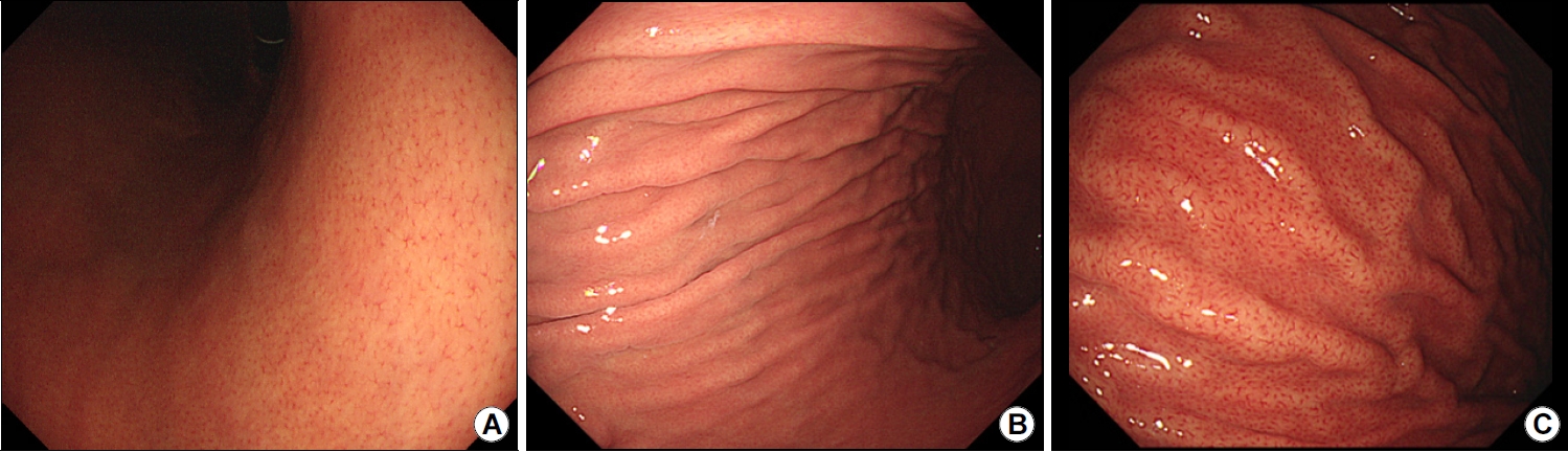
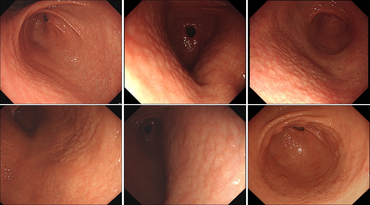
1) 위축

위축은 위 점막의 정상적인 위샘(gland) 조직이 지속적인 만성 염증으로 인해 소실되는 것이다. 위축의 내시경 소견은 점막의 전정부와 위체부의 소만 측에서 혈관 투영이 뚜렷하게 나타나고 위체부의 대만 측에서는 점막주름이 소실되어 보이는 것이다. 위축에 대한 내시경 소견으로 흔하게 사용하는 것은 Kimura–Takemoto 분류로 폐쇄형과 개방형으로 나뉜다(Fig. 3, 4). 위축은 일반적으로 폐쇄형에서 점차 개방형으로 진행하게 된다. 진료실에서 흔히 위가 얇아졌다라고 설명하기도 하는데 공기의 주입량이 많지 않았을 때 위의 점막이 얇아져서 점막하층의 혈관상이 보이는 위염이다. 공기의 주입량이 많을 때에는 개방형 위축처럼 보이기도 하니, 적절한 공기 주입이 필요하다. 또한 빈혈이 있을 때는 점막이 창백해지므로, 정상의 위의 경우도 위축으로 보일 수 있으니 빈혈이 있는 환자에서 위축의 진단은 신중하게 하여야 한다. 일반적으로 위축은 연속적으로 정상점막과의 경계를 찾아볼 수 있다. 국소적인 위축을 보이는 경우는 조직검사를 통하여 암을 감별하는 것이 필요하다(Fig. 5).
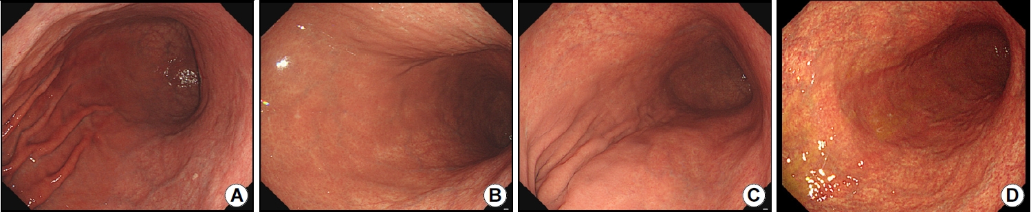
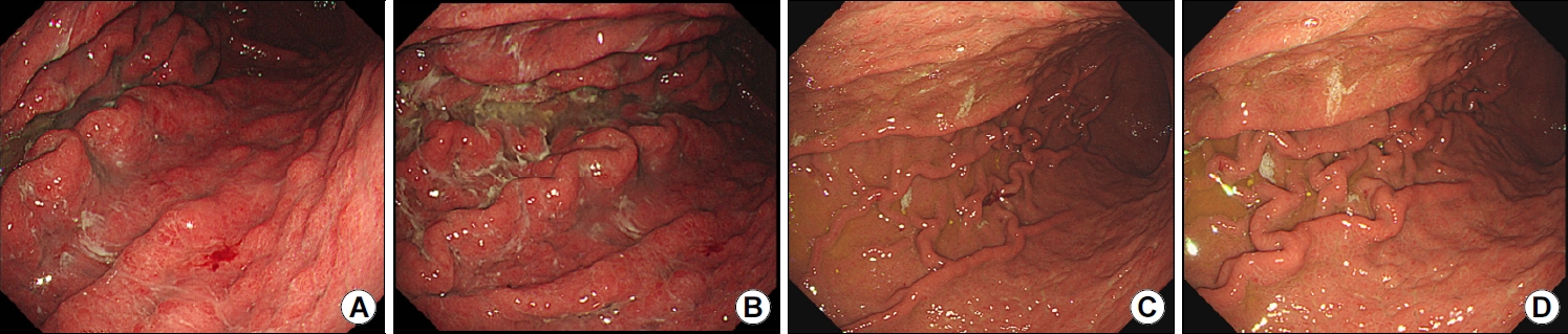
2) 장상피화생

내시경 소견은 워낙 다양한 형태를 나타내지만, 대표적인 것은 융기된 플라크 형태를 보이는 것이다. 회색 또는 흰색 점막의 융기된 병변이 단독으로 산재하기도 하고, 조약돌 모양으로 군집하게 되고 융합되는 소견을 보이기도 한다(Fig. 6). 흰색 또는 회색 점막, 거친 점막표면(rough mucosal surface), 융모성 모양(villous appearance)으로 표현하기도 하고, 군데군데 얼룩덜룩한 발적성 병변(mottled patchy erythema)으로 표현하기도 한다. 관찰자 간의 일치율이 낮아서, 메틸렌블루 또는 인디고카민 등의 색소내시경을 사용하기도 하나 연구적인 목적으로 사용하고, 진료환경에서 보편적으로 사용하기는 어려운 실정이다. 국내의 연구에서 백색광 내시경을 통한 장상피화생 진단의 특이도는 높으나 민감도는 상대적으로 낮았다[6]. 최근 내시경 기계의 발전으로 올림푸스사의 NBI (narrow band image), 후지논사의 FICE, 그리고 펜탁스의 i-scan이 임상에서 널리 사용되고 있다. 색소내시경보다 간편하기 때문에 임상에서 쉽게 이용할 수 있고, 협대역(NBI) 내시경으로 볼 때 백색광 단독으로 볼 때보다 융기된 점막을 쉽게 관찰하여 장상피화생의 진단을 높일 수 있다[7]. 포르투갈 연구에서 NBI 영상의 관융모점막(tubulo-villous mucosa) 소견으로 장상피화생의 높은 정확도를 보였다[8].
위축과 동반되는 경우가 많기 때문에 위축이 전정부에서 위체부 소만을 따라 진행되는 것처럼, 장상피화생도 전정부에 있는 경우와 전정부와 체부 모두 있는 경우로 구분한다. 내시경 결과지에 위암의 유병률이 높은 우리나라에서는 위축과 장상피화생의 정도를 내시경 결과지에 루틴으로 기록하는 것이 필요하다. 즉, 위축의 유무와 있다면 개방형인지 폐쇄형인지, 장상피화생의 유무와 있다면 전정부에 국한되어 있는 지 아니면 체부까지 있는 지를 기술하는 것이 좋겠다. 장상피화생의 유무도 중요하겠지만, 분포 또한 중요하다. 전정부에 국한되어 있는 경우보다 전체적으로 퍼져 있을 때 위암의 위험도가 높아진다[9]. 화생성위염은 위선종, 조기위암과 구별이 어렵기 때문에, 점막의 요철, 결손, 색조변화가 있다면 조직검사를 통해 정확한 진단을 해야 한다.
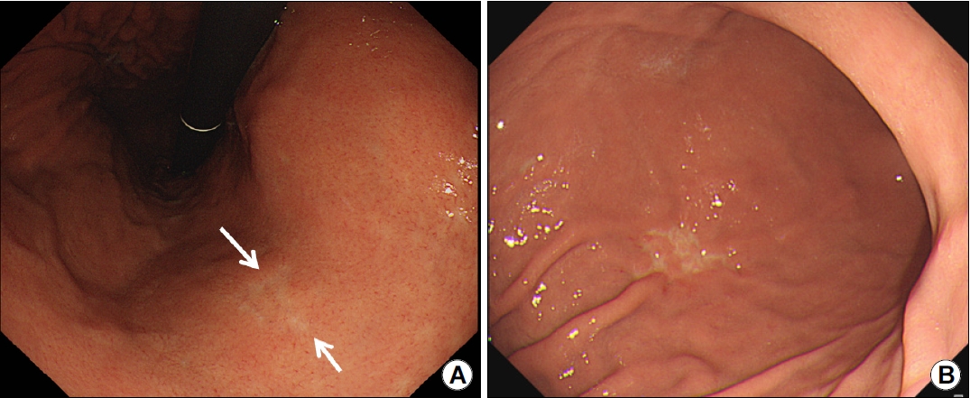
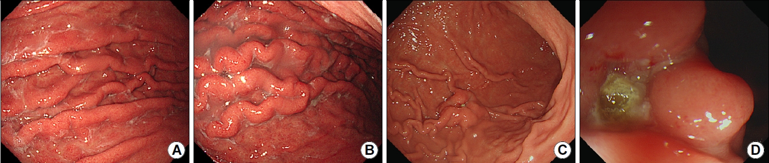
3) 미만성 발적

발적은 주로 체부와 기저부에서 관찰된다. 체부 전반에 널리 퍼져 있어 RAC 패턴의 관찰이 어려운 경우는 미만성(diffuse) 형태를 보인다. Kyoto 분류에서는 주로 체부에서 보이는 RAC를 기준으로 하여 RAC가 관찰되는 경우는 0점, RAC가 일부에서 관찰되면 1점, RAC가 소실된 경우를 2점으로 한다(Fig. 7). 이 소견이 H. pylori 관련 위염의 대표적인 소견이다[4]. 출혈반(hemorrhagic clot)의 소견은 발적과 다른데, 국소적인 형태를 주로 보인다(Fig. 8).

4) 결절

결절은 주로 전정부에서 관찰된다. 닭살 모양의 형태를 띠는 데 장상피화생의 내시경 소견과는 다르다. 결절은 크기가 균일하며, 작고 연속적으로 전정부에 분포되어 있는 데 반해, 장상피화생은 크기가 크고 다양하며 전정부 또는 체부까지 산재하여 있는 것이 특징이다(Fig. 9).
결절성 위염은 젊은 여자에게 흔하고 H. pylori와 연관이 있고 미만형위암으로 진행할 가능성이 있어 적극적인 H. pylori 치료가 필요하다. 치료 후 결절 또한 호전되는 것을 임상에서 경험할 수 있다[10].
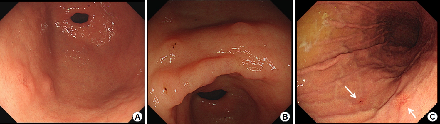
5) 주름 비대

주름 비대는 점막이 5 mm를 초과해 두꺼워져 있는 경우로, 보통 위 생검 겸자를 열었을 때 6 mm 정도 되기 때문에 그것 이상 두꺼워져 있다면 주름 비대로 볼 수 있다. H. pylori와 연관된 비후성위염과 보만4형위암을 반드시 감별해야 한다. 조직 생검에서 악성세포가 보이지 않고 H. pylori가 있다면 우선 H. pylori 치료를 한다. 주름 비대가 있는 H. pylori 양성 환자에서 H. pylori 치료 6개월 후에 주름 비대가 호전된 것을 확인할 수 있다(Fig. 10). 보만4형위암은 공기를 주입해도 위가 잘 펴지지 않는 특징이 있다. 대만의 주름을 잘 관찰하기 위해서는 충분히 공기를 주입하는 것이 중요한데, 주름의 종대가 국소적으로 있다면 얼핏 보기에는 국소적인 주름 비대로 생각할 수 있으나 점막 안에 숨겨진 종양이 있을 수 있기 때문에 보만2형 또는 3형 위암 여부를 반드시 확인해야 한다(Fig. 11).

4. 위축성위염과 장상피화생의 관리

환자들이 건강검진 결과표를 들고 와서 위축성위염과 장상피화생에 대한 질문이 진료실에서 가장 많이 듣는 질문 중 하나이다. 위축성위염은 나이에 따른 노화과정이고, 장상피화생은 위세포가 장세포로 바뀌는 것을 의미한다고 설명한다. 원래 장기 고유의 세포가 다른 장기의 세포로 바뀌는 것은 안 좋은 것이고, 나이가 들고 다른 기관이 노화가 진행하는 것처럼, 위도 노화가 되고, 그동안 몸에 안 좋은 담배, 짠 음식, 자극적인 음식 등을 먹어서 생긴 변화라고 환자에게 설명한다. 물론 시간의 변화에 따른 노화의 과정이기도 하지만, H. pylori의 존재에 따른 염증의 악화일 수도 있기 때문에 나이를 고려하여 판단하여야 하고, 젊은 사람에게 위축과 장상피화생이 나타난다면 H. pylori의 적극적인 검사와 치료가 필요하다.
위축성위염과 장상피화생의 임상적인 의의는 이형성을 거쳐 위 샘암에 이르게 하는 한가지 원인이 된다는 것이다. 외국의 연구에서 연간 위암발생률은 위축성위염 환자에서 연간 0.1%이고 장상피화생이 동반되었을 경우에 0.25%이다. 위암의 가족력이 있거나, 이전 시행 받은 위내시경 검사에서 위축성위염과 장상피화생이 동반되었을 경우에는 주의를 기울여야 한다. 위축성위염은 나이와 관계가 있다. 그러나, 70대에서도 상대적으로 폐쇄형을 보일 수가 있고, 40대에서도 개방형 위축성위염을 가질 수 있다. 이렇게, 생활습관의 관리가 중요한데 과음과 흡연을 삼가고, 짜고 뜨거운 음식을 포함한 자극적인 음식을 피하는 것이 도움이 되겠다. 특히, 소금과 짠 음식이 위암을 일으킬 수 있는 위험 요소로 분류되었고, 세계보건기구의 International Agency for Research on Cancer (IARC)는 소시지, 베이컨, 햄, 육포 등의 가공육 섭취가 위암을 일으킬 가능성이 높다고 하였다[11]. 또한, H. pylori가 있을 때 분문부 이외의 위암 발생 위험성이 증가했음을 대규모 전향적 영양 조사에서 보여주었다[12].
약물치료에 대해서 보면, 위축성위염과 장상피화생이 있는 환자에서 환자의 증상에 따라 약을 처방하지만, 환자들은 위염에 대한 약을 계속 먹으면 위염이 좋아질 수 있으리라는 희망으로 약을 복용하고 있지만 약물치료만으로 위염을 치료할 수는 없다. 다만, 다른 어떤 약보다도 H. pylori 치료를 하는 것이 좋은 선택일 수가 있다. 최근 연구에 의하면 H. pylori 치료를 함으로써 위축과 장상피화생이 좋아진다는 보고가 있어 H. pylori 감염이 있다면 제균 치료를 시행하여 위암 예방을 하는 것을 권유하고 있다[13,14]. 또한, 미국의 가이드라인에서도 루틴으로 내시경을 자주하거나 조직검사를 자주 하는 것을 권유 하고 있지 않지만, H. pylori 검사를 해서 H. pylori 치료를 하는 것은 권유하고 있다[9].
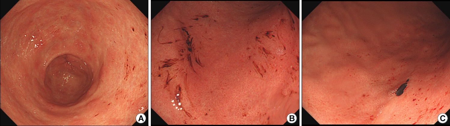
5. 미란

미란은 점막결손이 점막하층까지 닿지 않는 점막근판의 결손을 말하며, 점막근판을 침범하여 점막하층까지 결손이 있는 경우를 궤양이라고 한다. 점막근판 침범여부는 궤양의 크기로 유추하기 때문에 점막결손의 크기로 5 mm를 초과하면 궤양, 5 mm 미만이면 미란으로 한다. 미란은 융기형과 편평형으로 나눠서 기술한다(Fig. 12). 단독의 융기형 미란은 위선종, 또는 위암 가능성을 고려해서 조직검사가 필요하며, 다발성 융기형미란은 장상피화생과 혼동이 될 수 있다. 아스피린 또는 비스테로이드 항염증제(nonsteroidal anti-inflammatory drugs)는 미란 뿐 아니라 궤양을 일으키기도 한다. 주로 다발성 형태를 보이기 때문에, 다발성 미란 또는 궤양이 혼재해 있을 때에는 이런 약제를 복용했는 지 확인해야 하고, 보통 상복부통증 등의 증상이 있는 경우가 흔하다.

결 론

위염을 약으로 치료할 수 있다고 하는 것은 맞는 말일수도 있고 틀린 말일 수도 있다. 위염의 내시경 소견을 이해하고 H. pylori와 관련된 위염이라면 적극적으로 H. pylori 치료를 하고, 위염과 관련된 상복부통증과 속쓰림 등의 증상이 있다면 단기적으로 약물 치료를 한다. 단순히 내시경 결과지에 만성표재성위염이라는 용어와 진단을 사용하는 것보다는 위염의 Kyoto 분류에서 제시한 5가지 전암성 병변(위축, 장상피화생, 미만성 발적, 결절, 주름 비대)의 내시경 소견을 숙지하고, 체계적으로 내시경 소견을 기술하여 환자가 암에 걸릴 위험도를 평가하고[15], 적극적으로 H. pylori 제균 치료를 하는 것이 위염의 악화를 막고 위암을 예방하는 방법이다.

CONFLICTS OF INTEREST

There is no potential conflict of interest related to this work.

Fig. 1.
Endoscopic images showing regular arrangement of collecting venules across the gastric body as follows: (A) Along the lesser curvature. (B) Along the greater curvature. (C) In the high gastric body along the greater curvature.
kjhugr-2023-0012f1.jpg
Fig. 2.
Endoscopic images showing fundic gland polyps. (A) Multiple fundic gland polyps are observed in the gastric body. (B) A single fundic gland polyp is visualized at the cardia.
kjhugr-2023-0012f2.jpg
Fig. 3.
Endoscopic findings showing gastric atrophy. (A) Closed type 2 with atrophic changes involving the low gastric body along the LC. (B) Closed type 2 with normal RAC on the LC aspect of the high gastric body. (C) Closed type 3 with atrophic changes involving the gastric body along the LC. (D) Closed type 3 with normal RAC in the anterior and posterior walls of the gastric body. LC, lesser curvature; RAC, regular arrangement of collecting venules.
kjhugr-2023-0012f3.jpg
Fig. 4.
Endoscopic findings showing gastric atrophy. (A) Open type 2 with atrophic changes involving the anterior and posterior walls of the gastric body and normal RAC along the GC of the gastric body. (B, C) Open type 2 observed in the same patient. (B) Gastric folds disappear after insufflation of too much air. (C) Gastric folds can be visualized after slight suction of air. (D) Open type 3. GC, greater curvature; RAC, regular arrangement of collecting venules.
kjhugr-2023-0012f4.jpg
Fig. 5.
Endoscopic findings showing focal atrophy in the mid gastric body along the LC (A, arrow) and a discolored lesion involving the low gastric body along the GC. (B) Biopsy evaluation showing poorly cohesive carcinoma at both aforementioned sites of the gastric body. GC, greater curvature; LC, lesser curvature.
kjhugr-2023-0012f5.jpg
Fig. 6.
Endoscopic findings showing intestinal metaplasia. (A, B) Elevated white plaques (whitish granular plaques) are observed in the antrum (A) and at the gastric angle (B). (C, D) Elevated plaques are surrounded by a mixed patchy erythematous mucosal area in the antrum (C) and gastric body (D). (E, F) Gray plaques are visualized in the antrum (E); the plaques are more prominently observed using NBI (F). NBI, narrow band imaging.
kjhugr-2023-0012f6.jpg
Fig. 7.
Endoscopic finding of diffuse redness. (A) RAC. (B, C) Diffuse redness. Mild diffuse redness (B) and severe diffuse redness (C) involving the high gastric body along the GC. GC, greater curvature; RAC, regular arrangement of collecting venules.
kjhugr-2023-0012f7.jpg
Fig. 8.
Endoscopic image showing a hemorrhagic clot.
kjhugr-2023-0012f8.jpg
Fig. 9.
Endoscopic image showing nodularity. The “goose-flesh” appearance of the antrum is referred to as nodular gastritis or lymphofollicular gastritis.
kjhugr-2023-0012f9.jpg
Fig. 10.
Endoscopic finding of hypertrophic gastric folds. (A, B) Hypertrophy and mucosal exudates visualized in the gastric body along the GC. (C, D) Regression of hypertrophy is observed 6 months after Helicobacter pylori eradication therapy. GC, greater curvature.
kjhugr-2023-0012f10.jpg
Fig. 11.
Endoscopic findings of hypertrophic gastritis vs. Borrmann type 3 gastric cancer. (A, B) Hypertrophic gastritis. (C) Focal hypertrophic mucosa is observed in the low gastric body along the GC. (D) Cental ulceration of focal hypertrophic mucosa is visualized. GC, greater curvature.
kjhugr-2023-0012f11.jpg
Fig. 12.
Endoscopic images showing erosions. (A, B) Raised erosion. (C) Flat erosion (arrow).
kjhugr-2023-0012f12.jpg
Table 1.
Classification of Gastritis and Duodenitis Based on the Korean Standard Classification of Diseases (8th Version)
K29 Gastritis and duodenitis
K29.0 Acute haemorrhagic gastritis
K29.1 Other acute gastritis
K29.2 Alcoholic gastritis
K29.3 Chronic superficial gastritis
K29.4 Chronic atrophic gastritis
K29.5 Chronic gastritis, unspecified
K29.6 Other gastritis
K29.7 Gastritis, unspecified
K29.8 Duodenitis
K29.9 Gastroduodenitis, unspecified
Table 2.
Grading Scores Based on the Kyoto Classification of Gastritis
Parameter Score    
Gastric mucosal atrophy 0 None C0-CI according to Kimura-Takemoto classification
1 Mild CII-CIII
2 Severe OI-OIII
Intestinal metaplasia 0 None None
1 Mild Within the antrum
2 Severe Up to the corpus
Hypertrophy of gastric fold 0 Negative <5-mm gastric fold width
1 Positive ≥5-mm gastric fold width
Nodularity 0 Negative None
1 Positive Small nodules in the antrum
Diffuse redness 0 Negative None
1 Mild Mild translucency of collecting venules in the body
2 Severe Severe translucency of collecting venules in the body

Adopted from Sugimoto M, et al. Efficacy of the Kyoto classification of gastritis in identifying patients at high risk for gastric cancer. [3]

REFERENCES

1. Kim GH. Endoscopic findings of Kyoto classification of gastritis. Korean J Helicobacter Up Gastrointest Res 2019;19:88–93.
crossref pdf
2. Yagi K, Nakamura A, Sekine A. Characteristic endoscopic and magnified endoscopic findings in the normal stomach without Helicobacter pylori infection. J Gastroenterol Hepatol 2002;17:39–45.
crossref pmid
3. Sugimoto M, Ban H, Ichikawa H, et al. Efficacy of the Kyoto classification of gastritis in identifying patients at high risk for gastric cancer. Intern Med 2017;56:579–586.
crossref pmid pmc
4. Kang H, Lim CH, Kim S, Choi A, Oh JH. Usefulness of the Kyoto classification score for prediction of current Helicobacter pylori infection. Korean J Helicobacter Up Gastrointest Res 2022;22:281–287.
crossref pdf
5. Toyoshima O, Nishizawa T, Koike K. Endoscopic Kyoto classification of Helicobacter pylori infection and gastric cancer risk diagnosis. World J Gastroenterol 2020;26:466–477.
crossref pmid pmc
6. Lim JH, Kim N, Lee HS, et al. Correlation between endoscopic and histological diagnoses of gastric intestinal metaplasia. Gut Liver 2013;7:41–50.
crossref pmid pmc
7. Jeon SW. Endoscopic diagnosis and classification of atrophic gastritis and intestinal metaplasia. Korean J Helicobacter Up Gastrointest Res 2020;20:91–95.
crossref pdf
8. Pimentel-Nunes P, Dinis-Ribeiro M, Soares JB, et al. A multicenter validation of an endoscopic classification with narrow band imaging for gastric precancerous and cancerous lesions. Endoscopy 2012;44:236–246.
crossref pmid
9. Gupta S, Li D, El Serag HB, et al. AGA clinical practice guidelines on management of gastric intestinal metaplasia. Gastroenterology 2020;158:693–702.
crossref pmid pmc
10. Lee SY. Helicobacter pylori infection and the Kyoto classification of gastritis. Korean J Helicobacter Up Gastrointest Res 2019;19:81–87.
crossref pdf
11. Bouvard V, Loomis D, Guyton KZ, et al.; International Agency for Research on Cancer Monograph Working Group. Carcinogenicity of consumption of red and processed meat. Lancet Oncol 2015;16:1599–1600.
crossref pmid
12. González CA, Jakszyn P, Pera G, et al. Meat intake and risk of stomach and esophageal adenocarcinoma within the European Prospective Investigation Into Cancer and Nutrition (EPIC). J Natl Cancer Inst 2006;98:345–354.
crossref pmid
13. Kim BJ. Natural course of atrophic gastritis and intestinal metaplasia. Korean J Helicobacter Up Gastrointest Res 2020;20:101–106.
crossref pdf
14. Lee Y, Jeon YC, Koo TY, et al. Histological changes of gastric atrophy and intestinal metaplasia after Helicobacter pylori eradication. Korean J Gastroenterol 2007;50:299–305.
pmid
15. Lee SY. Association between the Kyoto classification of gastritis and the updated Sydney system. Korean J Helicobacter Up Gastrointest Res 2022;22:166–169.
crossref pdf


Editorial Office
Lotte Gold Rose II Room 917, 31 Seolleung-ro 86-gil, Gangnam-gu, Seoul 06193, Korea
Tel: +82-2-717-5543    Fax: +82-2-565-9947    E-mail: helicojournal@smileml.com                

Copyright © 2024 by Korean College of Helicobacter and Upper Gastrointestinal Research.

Developed in M2PI

Close layer
prev next